Archive for the ‘scan’ tag
3DsMax From Home
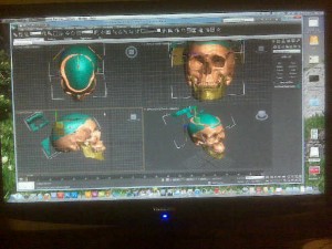
This weekend I installed the Autodesk Education Suite for Entertainment Creation 2011 into my new desktop computer. There may still be a couple things to sort out, but overall, my office has taken a right leap in terms of capabilities over these past few weeks. Just tonight I brought in the .stl files I have been working on at school with Mimics. There they are!
To get these, we ran a cat scan on a plastic skull model over at the craniofacial clinic. The dicom data was then put onto disk and taken back to the biomedical visualization computer lab. There I imported the files into Mimics and began the tedious process of sorting out the wanted bits from the unwanted bits, and deciphering layer by layer the mandible from the whole of the skull. I have to say, the teeth were tricky to get right, but it looks like it came out pretty well.
Once the Mimics files were set up, I was able to create 3D models and export .stl files and save them to a thumb drive. Tonight I imported those .stl files into my very own copy of 3DsMax and began sorting through extraneous polygons. If you haven’t worked with these programs before, you might think of this as the 3D version of cleaning up the edges and unwanted specks from everything. I still have quite a bit more of this to do.
I also got everything lined up. The mandible and majority of the skull were from the same cat scan so they imported in perfect alignment, but the calvarium was done separately on account of the I-Cat not being large enough to scan the whole skull in one take (it is mostly used for dental purposes over there).
So far so good, and I’m looking forward to working with this model for my upcoming animation. I decided to go with the plastic model in the end, rather than the actual patient data because it is more normalized, and I can alter it to create the pathology I want to focus on without a lot of other distracting anomalies getting in the way. Actual people rarely come so standardized, especially the ones who are coming in for craniofacial surgery. Plus I am able to get a much cleaner scan because there is no soft tissue to weed out of the data, and I’m also able to get cleaner teeth without distortion from metal braces, which any patient preparing for the procedure I will be animating would be wearing.
All in all, I think this is the best way to go, and I’m excited to get to start working on this from home now, rather than trying to remember the best hours to work in the computer lab at school. And oh, after so much work in Maya recently, I’m surprised at how nice it is opening up 3DsMax again. Yay.
A Pin-Up Calendar That Shows All
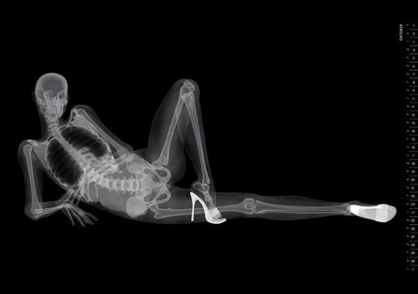
Y’know I just keep coming across this, and well, I thought this time that I would share it here.
http://www.butter.de/fileadmin/kampagnen/eizo/eizo_kalender.swf
It would seem that the medical imaging company, Eizo, has teamed up with ad agency Butter to create a truly unique pin up calendar. All I can say is congratulations guys, because this promotion is certainly making the rounds. And as well it should be! What a fun idea!
Art from a Scan
Sometimes people send me things; links, images, stories, things related to medical art.
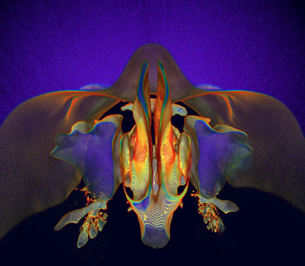
Today someone sent me the winning image of the 2007 International Science and Engineering Visualization Challenge, entitled “What lies behind our nose” created by Kai-hung Fung.
This is a great example of the kinds of things that can be done when scanning technology meets art. The image started as a CT scan from a 33 year old woman, who was being examined for thyroid disease.
I’m not sure what kind of software Fung used here, but if you want to experiment with this kind of art a little yourself, you might start with a freeware copy of OsirX.
http://www.osirix-viewer.com/AboutOsiriX.html
(Sorry PC users, but this one is Mac only)
From there, you have some options regarding what kind of tissue you want to select for, how colors are displayed, as well as the ability to cut pieces away, or turn your model. It takes some getting used to, and I am by no means an expert with this one yet, but the results can be downright cool. It’s also just a nice way to be able to view DICOM images on your own computer. If you don’t have access to any DICOM images, and you still want to try some of this first hand, you can always download one of the sample image sets available.
http://pubimage.hcuge.ch:8080/
You can read more about Kai-hung Fung’s piece, as well as the other 2007 Visualization Challenge winners here.
http://www.sciencemag.org/cgi/content/full/317/5846/1858


