Archive for the ‘muscle’ tag
Animation Finished!
This week I finally finished the 3D animation that I have been working on this semester. It’s subject is the sliding filament theory of muscle contraction and I believe that I have managed to put together something clear.
I find that I still struggle in places with 3DsMax, but learning After Effects and coming back to working in Final Cut Pro again was a lot of fun. Our class on the whole created some really great work.
Science Photo Library
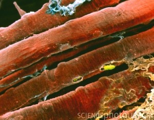
So I was tracking down the image below, and discovered a new resource for good scientific images.
The website it came from is http://www.sciencephoto.com/. The image itself is credited to Professors P.M. Motta, P.M. Andrews, K.R. Porter “& J. Vial. Apparently, this is what one can do with an electron micgrograph and some skill with color. This is actually the shot that first made me understand that there are two kinds of striation in skeletal muscle, the actual fibers, and then the banding created by the myofilaments arranged inside. Take a close look!
The Scapula
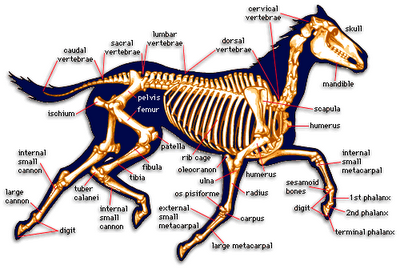
Lately, I’ve been thinking a lot about the scapula. It’s such a uniquely shaped bone, and such a central point to so many muscles. It is fundamental to the shoulder, but there isn’t anything like it in the hip. I suppose that one could argue that the shape of the pelvis accounts for the needs of the lower limb, and perhaps it is useful to have a more solid skeletal arrangement in our weight bearing joints, but then why do horses have scapulas?
I suppose it must be some kind of evolutionary remnant. Perhaps we’re all meant to have wings. Or I suppose it could just be the way things have come together around the ribcage.
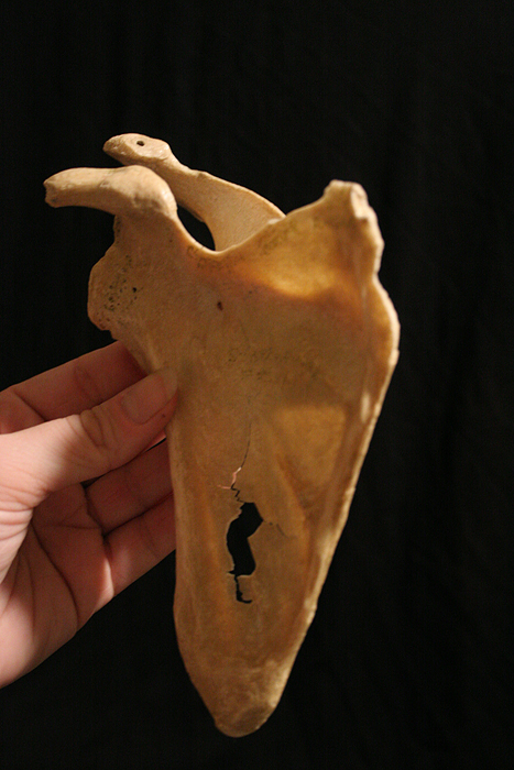
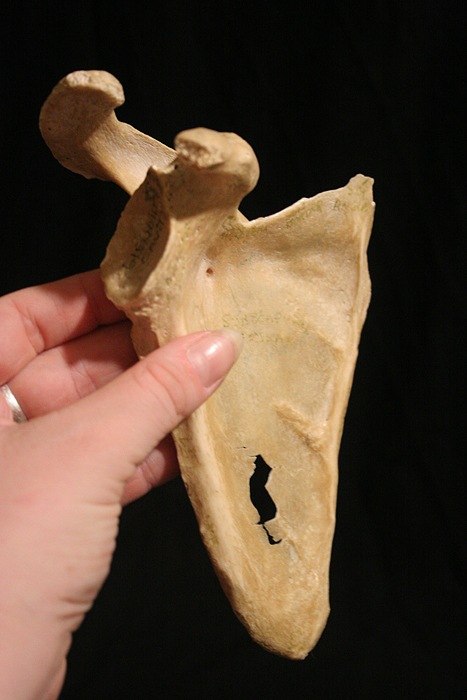
But musings aside, there is a lot to know about this bone. The photo above is taken from an anteromedial angle, so the protruding hook like shape up top is the coracoid process, and the curve in back is the acromion. The acromion comes out of what is considered the spine of the scapula. Between those two processes there is a space that runs the width of the scapula called the supraspinous fossa. The glenoid cavity is particularly important as it creates the glenohumeral joint with the humerus. It can be better viewed from the anterolateral view in the picture below. The bulges of bone matter on the top and bottom of that glenoid cavity make up the supraglenoid tubercle and the infraglenoid tubercle.
There are a lot of muscles which attach to the scapula. You wouldn’t think there would be room.
Supraspinitus sits in the supraspinitus fossa up top. It is the first of the SITS muscles (or rotator cuff muscles), which all plug in to the humerus distally from the scapula. It is innervated by the suprascapular nerve which travels through the suprascapular notch (sometimes called the spinoglenoid notch) in the scapula which occurs near the base of the coracoid process. The popular mneumonic “army over navy” is meant to describe the relationship between the suprascapular artery and nerve, as the artery (army) goes over the suprascapular ligament, where as the nerve (navy) goes under said ligament and through the notch.
Infraspinitus is the second in the SITS muscles. It attaches at the infraspinous fossa of the scapula (that would be the big flat part on the dorsal side) and reaches over to the greater tubercle of the humerus. It, like the supraspinitus, is inervated by the suprascapular nerve, and it is supplied by both the suprascapular and circumflex scapular arteries.
Teres minor would be the T in SITS, and it attaches to the scapula at the lateral border below infraspinitus and like the first two muscles here, attaches to the greater tubercle of the humerus. It’s innervation comes from the axillary nerve, and it’s blood supply comes from both the posterior circumflex humeral artery (which you may recall if you’re studying this stuff, travels along with the axillary nerve through the quadrangular space), and the circumflex scapular artery, which branches off the subscapular artery (and courses through the triangular interval) to form an anastamosis around the scapula with the suprascapular artery that I just listed as supplying the two superior muscles here.
And lastly, in the SITS mneumonic we have subscapularis. Subscapularis is the inferiormost muscle in SITS and it attaches to the scapula at the subscapular fossa (big flat area on the ventral side) and reaches over to the humerus, this time attaching at the lesser tubercle. It is innervated by the superior and inferior subscapular nerves. I have disagreeing sources as to where it’s blood supply comes from, but the subscapular artery is definitely involved.
Since we discussed Teres Minor, it only makes sense to move on to Teres Major. Teres Major takes it’s place on the inferior angle of the scapula. So it has the most inferior attachment on the scapula of any muscle. It also attaches on the humerus, but further down on the medial lip of the intertubercular sulcus (otherwise known as the bicipital groove). It is mostly innervated by the lower subscapular nerve, and supplied by the subscapular and circumflex scapular arteries.
Deltoid wraps over all of those mentioned humeral attachments and comes up to attach to the spine of the scapula, as well as the acromion, and also the lateral third of the clavicle. Then distally it attaches to the deltoid tuberosity of the humerus. It’s innervation and blood supply come from the axillary nerve and posterior circumflex humeral artery, which have traveled together through the quadrangular space to get to it, stopping breifly at Teres Minor on the way.
Trapezius, along with Deltoid, are the two most superficial and form defining muscles attached to the scapula. Actually, Trapezius is huge and attaches all over the place, but on the scapula it attaches at the acromion and spine, superiorly to the Deltoid. It is innervated by the spinal accessory nerve (CNXI) and receives blood from the transverse cervical artery.
Then, just beneath your Trapezius you’ll find the rhomboids, both major and minor. They reach from the spinous processes of vertebra C7-T5 (the distinction between the two coming between T1 and T2) to the medial border of the scapular spine. As usual, when you have a major and a minor of a muscle, the minor sits on top and is smaller than the major. Both are innervated by the dorsal scapular nerve, and supplied by the dorsal scapular artery.
Levator Scapulae is also innervated and supplied by the dorsal scapular nerve and artery, though it also takes some innervation directly from spinal nerves C3 and C4. It is positioned just superior to the rhomboids on the medial border of the scapula, which puts it just anterior to the scapular spine. And it comes from all the way up the spine at transverse processes of C1-C4. As the name would indicate, levator scapulae elevates the scapula. It is often called the “shrugging muscle.”
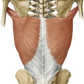
And while we are looking at the back, I may as well jump back down the spine to mention latissimus dorsi, aka “the climbing muscle.” This one only barely catches the inferior angle of the scapula and honestly, I have yet to see a picture that represents it as connecting at all, nor have I particularly noticed it in my own dissections (though to be fair, I wasn’t exactly looking for it). It has a much more noticable attachment on the spinous processes of C7-T1 as well as the illiac crest of the pelvis, inferior ribs and most distally at the floor of the intertubercular sulcus on the humerus after twisting around teres major. If you look closely, you can kind of see what that looks like here (taken from Thiemeteachingassistant.com).
Latissimus dorsi is innervated by the thoracodorsal nerve which comes off the posterior cord, and supplied by the thoracodorsal branch off the subscapular artery.
Then anteriorly, coming from the medial costal surface of the scapula to the first eight ribs you’ll find serratus anterior. Attaching in bundles to each of those eight ribs is what gives the muscle its serrated appearance, and therefore name. It is also known as the “boxer’s muscle” because it gets worked when someone throws a punch. Serratus anterior is innervated by the long thoracic nerve, which is one of the more easily damaged nerves because of it’s long unprotected journey along the superficial thorax. And it takes blood from the lateral thoracic artery (Wikipedia includes a second arterial supply as well, but I have not seen that artery listed in other sources.)
Near the serratus anterior, you will find the pectoralis minor. Only minor attaches to the scapula, pectoralis major goes over pectoralis minor to attach at the clavicle and sternum. But pectoralis minor attaches right onto the coracoid process of the scapula, and reaches inferiorly to ribs 3-5 near the costal cartilages. It is innervated by the medial pectoral nerve, and usually the lateral pectoral nerve as well. And it is supplied with blood by the pectoral branches of the thoracoacromial trunk.
Coracobrachialis also attaches at that coracoid process on the scapula (as the name would suggest). It is the smallest of the three muscles that attach there, and I’m a personal fan of it because it makes for an easy orientation point when you’re trying to sort out the brachial plexus. You can always spot coracobrachialis because it is pierced by the musculocutaneous nerve, and you can always spot the musculocutaneous nerve, because it is the only one piercing a muscle like that in that axillary area. And yes, the musculocutaneous nerve is the one innervating this muscle.
Then the third muscle attaching to the coracoid process is the biceps brachii, specifically, the short head of the biceps brachii. The long head runs through the intertubercular (or bicipital) groove of the humerus and attaches at the supraglenoid tubercle of the scapula. Both heads distally attach at the tuberosity of the radius and bicipital aponeurosis. The biceps brachii are also innervated by the musculocutaneous nerve, just after it comes out the other side of coracobrachialis. It is the continuing fibers of the musculocutaneous nerve after they have passed through the biceps (around the elbow) that are then referred to as the lateral antebrachial cutaneous nerve.
And opposing the biceps brachii, there are the triceps brachii muscle (hardly sounds like a fair fight now does it?) It is only the long head of the triceps brachii that attaches to the scaplula. This attachment occurs at the infraglenoid tubercle. The lateral and medial heads attach at the posterior humerus, superiorly and inferiorly to the radial groove respectively. Distally, all the heads attach at the olecranon process of the ulna. Since the axillary nerve already broke off and went up to innervate teres minor and deltoid, the only branch left off the posterior cord to supply another posterior compartmentalized muscle is the radial nerve, and that’s where triceps brachii gets it’s innervation. And it’s blood supply comes from the deep brachial artery (otherwise known as profunda brachii.)
Now if you’re studying the shoulder area, you can count yourself done, but I would be remiss if I didn’t after all this admit that the omohyoid muscle does in fact also attach to the scapula. It rises from the superior border, or sometimes from the suprascapular ligament and inserts on the hyoid bone in the throat.
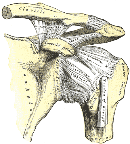
So as you can see, that’s a whole lot of muscle pulling that bone all around. And I know I only mentioned the suprascapular ligament specifically (which I should say also goes by the name superior transverse scapular ligament), but there are also the coracoclavicular ligaments, an acromioclavicular ligament, a coracoacromial ligament, a coracohumeral ligament, and a whole coating over the glenohumeral joint called the joint capsule glenohumeral ligaments and the axillary recess (at the base of that joint, my guess is that the axillary recess is there to provide a little extra material and enable more movement in the joint.) Here is the old Grey’s Anatomy image (grabbed from Wikipedia) to help illustrate those, though if you pay close attention to the names, you will find them mostly clearly named for the structures they connect.
So yeah, there’s a lot going on in here. As a friend recently said “No wonder it took 2.5 years for my shoulder injury to heal.”
Illustrating Muscle Contraction – Many Ways to Tell the Same Story
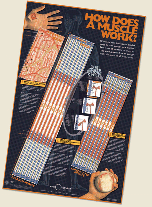
Earlier this week, I wrote about 2007 Visualization Challenge winner, Kai-hung Fung. I couldn’t help but notice one of the honorable mentions went to artists, Mark McGowan, Pat Murphy, David Goodsell and Leana Rosetti for their poster “How Does a Muscle Work.”
It caught my eye primarily because we were assigned the same process to illustrate our first semester in the biomedical visualization program at UIC.
The process of muscle contraction is actually pretty complicated. It is best understood when looked at on both the molecular level, but then also on the level of the actual sliding filaments in order to understand what exactly is accomplished by that molecular action.
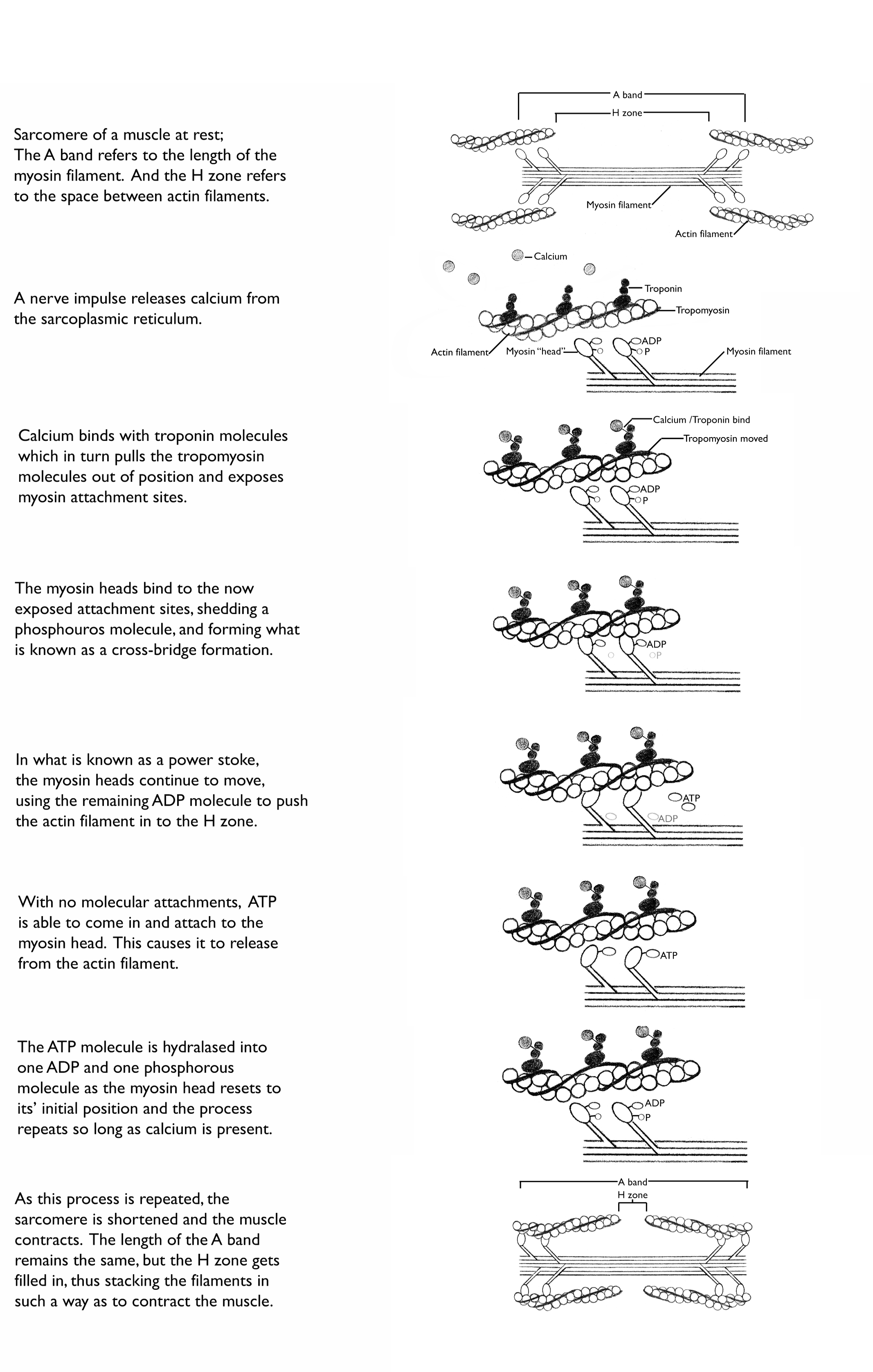
My own take on this process for the class, was to use very simple illustrations and explain the steps, with the larger view of the sarcomere at the beginning and end of the process.
Other classmates went about illustrating the process very differently.
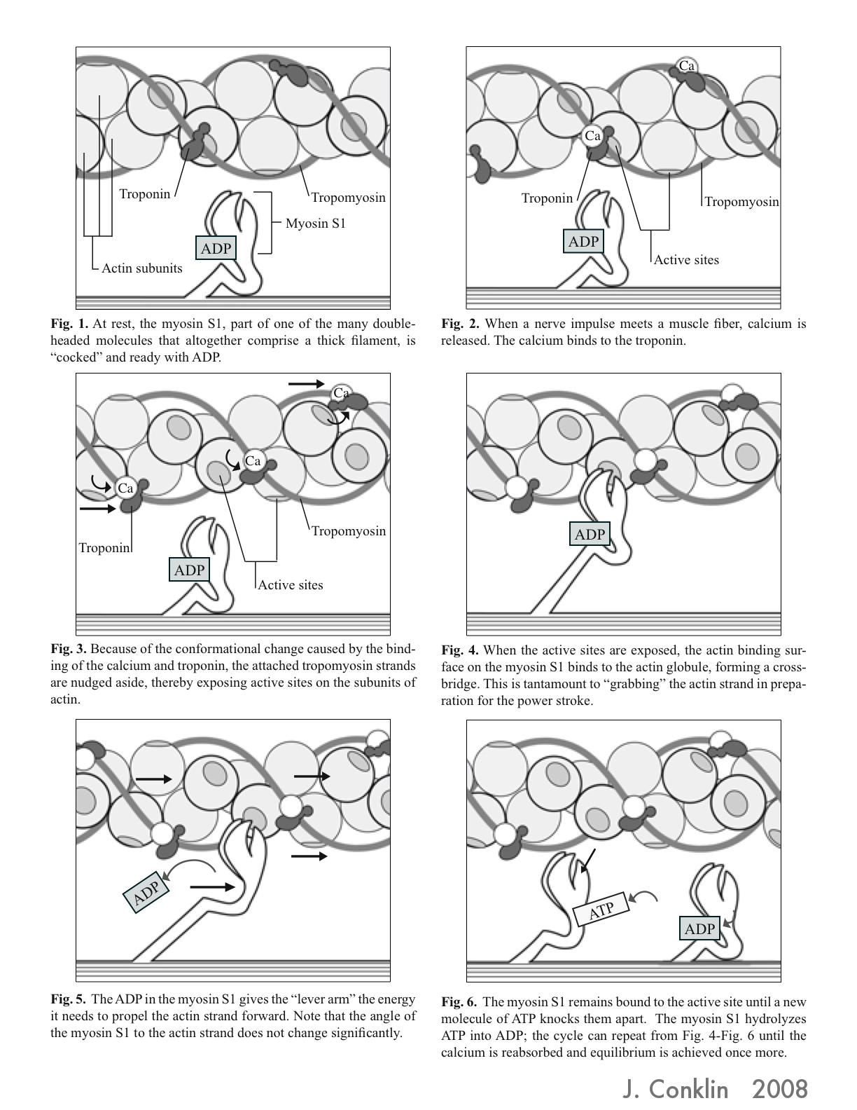
One of my favorites was that of Josy Conklin, who went out of her way to learn the real shape of the myosin head and bring that information into her illustration.
She posts this illustration to her own blog, Josydoodle, which I recommend to anyone who enjoys reading about these kinds of things.
http://josydoodle.blogspot.com/2008/12/myosin-yourosin-hisosin-herosin.html
Other classmates included less of the explaining text, and focused more on dynamic imagery. Some represented the power stroke cycle circularly, to emphasize the repetition involved in the process.
As you can see though, there are many different ways to tell this story. And that, I would say, is one of the primary issues a medical illustrator faces. A medical illustrator not only has to understand the science to be conveyed themselves, but they then have to find the best way to convey that information to the audience at hand, and utilize the media tools available to the specific project. And as with any story, there are many ways one can tell it.
