Science Photo Library
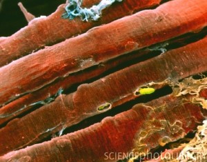
So I was tracking down the image below, and discovered a new resource for good scientific images.
The website it came from is http://www.sciencephoto.com/. The image itself is credited to Professors P.M. Motta, P.M. Andrews, K.R. Porter “& J. Vial. Apparently, this is what one can do with an electron micgrograph and some skill with color. This is actually the shot that first made me understand that there are two kinds of striation in skeletal muscle, the actual fibers, and then the banding created by the myofilaments arranged inside. Take a close look!
