Archive for the ‘arteries’ tag
Through a Blood Vessel
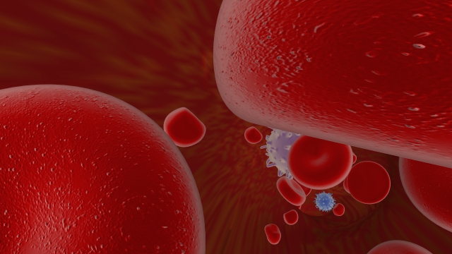
Last week I finally managed to export a playblast from Maya of my blood vessel project. It’s nothing too impressive at this stage, but I’m liking the roller coaster ride feeling of it…
Here are a few stills to give you a better idea of what the final render will look like…
I recently decided to end the ride with the blood cells being pulled off course up into a blood clot. Seemed like a fitting end to the journey. But I’m currently having some difficulty with rendering images that use a jpeg in the color channel. I’m still not certain if this is a matter of my laptop’s capabillity or some limitation of the Maya Hardware renderer which has allowed me to do some other tricks with my textures that I really don’t want to give up. Anyway, here is a screen shot of that final clot area as it stands now.
I still need to model some fibrin in there to really say blood clot, but the rendering difficulties have made it very difficult to do anything lately. I’m trying to change up the texture of the vessel walls themselves as well. Fingers crossed for awesome results!
Legs Legs Legs!
Ah yes, so I’ve been studying legs tonight. And I did a little sketching to help me get it all straight.
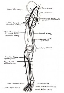 All together, I believe this is the gist of the situation.
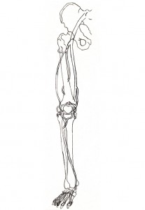 Here is the same thing with just the arteries.
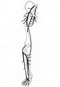 And here it is again with just the nerves. Those dots at the end represent the various muscles being innervated. They include iliacus, sartorius, recuts femoris, and your three vastus muscles off the femoral nerve. Then pectineus, obturator externus, gracilis, adductor magnus, adductor brevis, and adductor longus off the obturator nerve. And then the fibularis longus and fibularis brevis off the superficial fibular nerve, and extensor digitorum longus, tibialis anterior, extensor hallucis longus, fibularis tertius, and extensor digitorum brevis off the deep fibular nerve.
A few neurovascular relationships worth noting are that the femoral triangle contains the femoral nerve, artery, and vein, in that order going laterally to medially. The great saphenous vein (not pictured here) accompanies the saphenous nerve. And in the back (and thus not visible here) the small saphenous vein accompanies the sural nerve.
The above drawings are from an anterior view, with some reference as to what’s going on in back so long as it doesn’t cross behind bone. Do not use these for reference as to whether the nerve or artery is superficial or deep. Both systems are fully drawn without regard for empty spots where one crosses in front of the other. And lastly, I really wish I could give the photographer credit for the first image up there, but alas I cannot make out what the watermark says. But thank you G.S. for such a beautiful image, and good reference for leg and foot surface anatomy!
Blood Flow Out Into The Arm
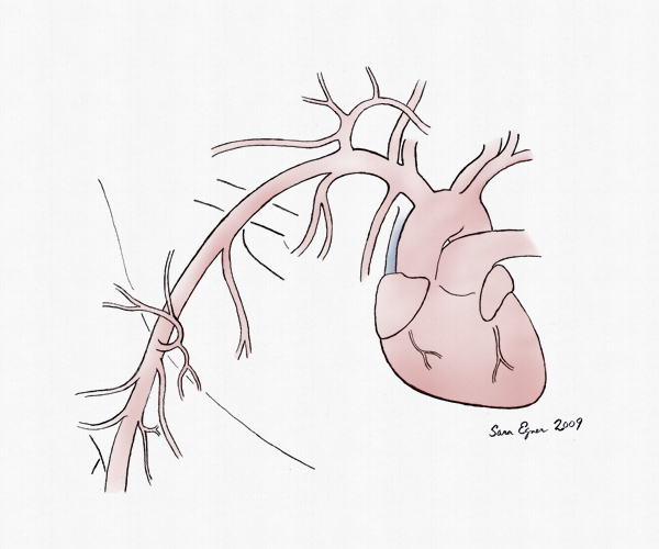
Blood gets pumped from the left ventricle, out into the aorta, where it goes either into the brachiocephalic trunk to get to the subclavian artery if it’s going to the right limb, or directly into the subclavian artery if it’s going to the left limb.
From the subclavian artery there is a branch to the vertebral artery, the internal thoracic artery, and the thyrocervical trunk, which has several branches including the suprascapular artery (which gets seen in an axillary dissection at the point where it courses under the suprascapular ligament and goes through the suprascapular notch) and also the transverse cervical artery (which supplies trapezius with it’s superficial branch, and if it splits into a deep branch supplies latissimus dorsi, levalor scapulae, the rhomboids, and more of trapezius).
Then costocervical trunk comes off of what is known as the second part of the subclavian artery. It branches into a Deep cervical artery, and a supreme intercostal artery.
The third part of the subclavian artery usually has no branches. But in people who’s transverse cervical arteries don’t split into a superficial and deep branch, you will find the replacement for that deep branch here. In which case it will be called the dorsal scapular artery and do all the things described above as the deep branch of the transverse cervical artery.
Then once you’ve passed by the first rib, the subclavian artery becomes known as the axillary artery. There is a popular mnemonic for this part, and it goes Suzi Taylor likes pot and sex (or sex and pot, those last few don’t always line up just perfectly). So that all goes…
Suzi – Superior (or Supreme) thoracic artery, which supplies the upper thoracic wall
Taylor – Thoracoacromial artery, which branches into an acromial, clavicular, deltoid, and two pectoral branches (you can spot it ‘cuz it looks like a little tuft of arteries bursting out there)
Likes – Lateral thoracic artery, which supplies the lateral thoracic wall
Pot – Posterior circumflex humeral artery (which then travels with the axillary nerve through the quadrangular space – I like to think of Ax smoking pot while he goes through a square environment)
And – Anterior circumflex humeral artery (which is smaller that the posterior one)
Sex – Subscapular artery, which immediately divides into the circumflex scapular artery and thoracodorsal artery. I’ve labeled this one lastly here, mostly to match the drawing above, but it often comes out before the humeral ones. you’ll know it by being the one in that last bundle before getting to Teres Minor where there is an immediate branch, and also because it will come off medially toward the scapula as opposed to the circumflex humeral arteries which both go wrapping around the humerus.
After the border of Teres Minor, the major artery becomes known as the brachial artery. I haven’t drawn that part here, but it will give off the profunda brachii, superior and posterior collateral arteries, and then break into a radial artery and an ulnar artery somewhere around the cubital fossa.

