Archive for March 21st, 2018
Nephron Anatomy
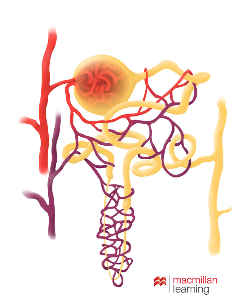
I recently had an assignment to create a nephron illustration. I’ve illustrated nephrons before, and I even thought I’d be able to re-use some of my old work. But it turns out, all of my previous work with nephrons was at a more introductory level. I have to say, I was downright disappointed. Because science gets complicated, and anatomy in particular, there are a lot of times when models are simplified for clarity, but it really bothers me when I feel like students are actively mislead.
The first time I ever illustrated a nephron was in grad school. I did it for my pen and ink assignment, and was really happy with how it turned out.

Then not long after I started with Sapling Learning, I wound up illustrating a nephron in Photoshop.

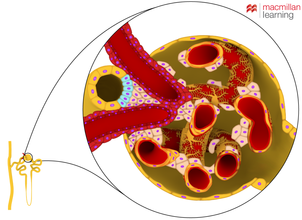
We even used arrows with this one at one point to show flow. And I looked at a lot of images throughout making both of these images. But not until a more advanced illustration request came up, did I ever realize that the distal tube of the nephron always passes by the opening of the capsule between the efferent and afferent arterioles. There’s actually an important feedback process that happens there where the contents of the tubule affect dilation and therefore the rate of filtration happening.
If you look up the juxtaglomerular apparatus you can find a lot of references at this level. That one detail of form is an important part of how nephrons work. So it bothers me that it’s so widely agreed not to feature it in more introductory images. They do that for clarity, but I think it’s too important and really I’m not sure that it actually adds all that much more complexity. So this is my most recent nephron artwork. It’s not my prettiest version, but it does tell more of the story. If we’d had more time, we probably would have done more with the form of the podocytes. They really weren’t the point of the illustration though, so we settled for at least giving them a nod. This was more about showing the layout of the cells between where the distal tubule passes between those in and outgoing arterioles to allow for the signal exchange about how much filtration is needed and for flow to be appropriately affected via dilation.

So now I’m hoping that moving forward, I can always use that simple design change in representing nephrons so that when students get to this part, it’s not so jarring. Maybe a lot of students don’t get stuck on details like that, but I know that I always have, and I know that I’m not the only one.