Archive for the ‘photography’ tag
Miru Kim
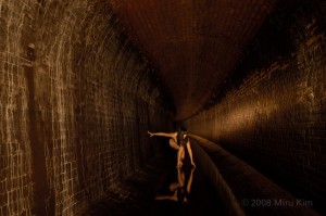
I was scoping around the internet a bit today for interesting anatomy and art related projects when I stumbled across photographer Miru Kim. Miru Kim is not an anatomical or medical artist, but rather she is an artist who takes some of her inspiration from early anatomy lessons. Her work deals more with what’s under the skin of a city, and urban decay, but I was so impressed with her that I simply had to write about her here.
Playing both the role of photographer and model, she has captured photos for her series, Naked City Spleen, across various cities spanning several countries, and getting into fascinating underground and decaying places. I am usually the last to be impressed by themes or series in an artist’s work, but there is something about the repeated juxtaposition of her naked figure against these decaying environments that speaks to the vulnerable nature of humanity and yet at the same time emphasizes a strength and bravery as you begin to contemplate the positions that this artist has put herself into for her art.
Below I’ve embedded the TED talk she gave in 2008. It is well worth the watch.
Microscopic Photography
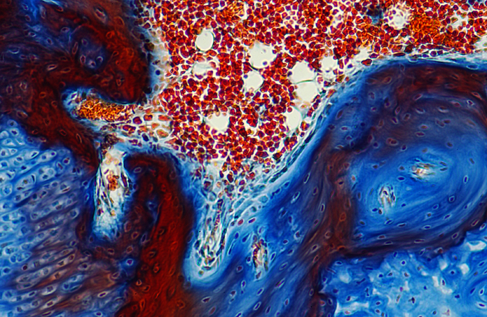
Today I got to take pictures with a microscope. I’d been given the overview of how it all worked once before, but today was the first time I ever really got to play with one. Everything I shot was off of the same slide. I’m told it was a mouse cochlea, but it looked like art to me.
This one was my favorite image of the bunch…
The rest can be seen here…
http://snapshotgenius.com/gallery/rodentcochlea
These were fun to take!
note: further conversation with the histologist who stained the slides in the first place, Dr. TK Bhattacharyya, revealed this to actually be the cochlea of a guinea pig. Live and learn.
Surgical Media
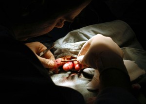
Yesterday I met with a very interesting man on campus, Chet Childs, about doing an independent study with him. The original thought was to learn about medical and scientific photography from him, but it looks like he’s doing a lot of surgical video work as well, which I have also been interested in for quite some time. I have a strong background in both photography and video editing. These were my primary means of income when I lived in LA. So finding someone who is using those kinds of skills in this, my new world of medicine and anatomy is such a perfect move for me. Sadly, I hear this is a dying niche, budget cuts, and these hard economic times, and all those lines we’ve all heard so very many times at this point. But there seems to be a need. So I’m going to look into this.
photo by Dexter Roberts
The Bassett Collection
Ok, so this is I guess the part where I wonder what kind of audience I’m writing for here. Many of you, will already know about this one, but for those of you that don’t, let me introduce you to the Bassett collection. I first stumbled upon these works through my school. My first class with cadavers prompted a near instant interest in cadaver photography in me. I’m still not sure whether the recommendation was meant to inspire me, or to show me that it had already been done and done well (there are a lot of both legal and ethical issues involved in cadaver photography), but I was told about some truly great work.
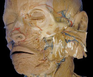
Beautifully lit, so clear, and at times haunting, this collection has to be the greatest of it’s kind out there. The shots are actually taken in 3-D and if you can get yourself a copy of the Stereoscopic Atlas of Human Anatomy, and any simple 3-D viewer (that’s right, the kind you had when you were a kid), you too can view the reels in all their 3-D glory.
As you can see here though, even the 2-D versions of these images are stunning, and well worth a look.
The collection resides at Stanford University’s School of Medicine. It came about as a 17 year collaboration between David L. Bassett, and photographer William Gruber.
*and FYI – Gruber actually invented that little plastic Viewmaster you had as a child!*
To view Stanford’s collection online, go here…
http://lane.stanford.edu/bassett/index.html
